Ultrasound is the primary imaging modality for evaluating the testes and surrounding tissue. The most common symptom will be a palpable lump felt in the testes. Other reasons for a testes scan include pain, undescended testes, inflammation, trauma, follow up on varicocele and hydrocele diagnosis.
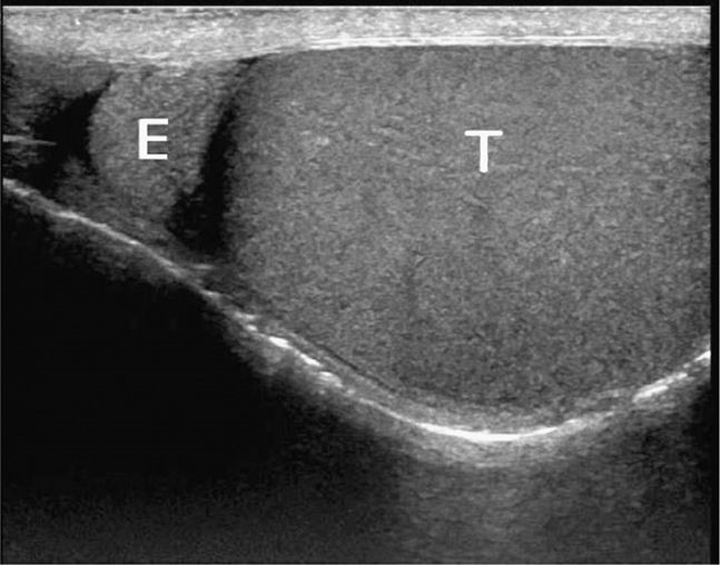
Ultrasound images of a normal testes

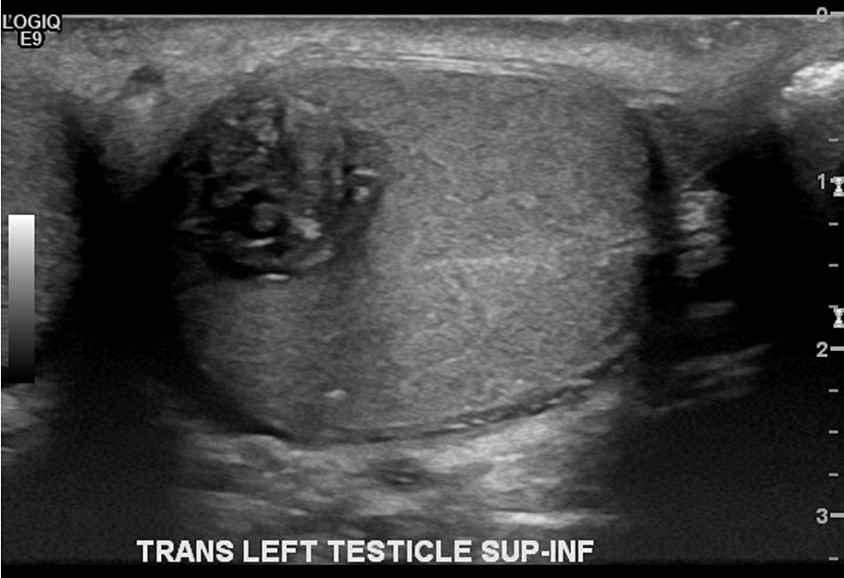
Ultrasond of an abnormal testes (testicular lump)

Preparation and Ultrasound examination
No specific preparation is required prior the Ultrasound examination. Before the scan, verbal consent is sought and you will be asked to undress from waist down and to lie on your back with towels covering you. Ultrasound gel is applied on the testes and an ultrasound probe (camera) is used to move around testes and surrounding tissue whilst images are acquired. This procedure should take approximately 15 minutes.
After the examination you will be given towels to remove the gel and dress up. The Sonography practitioner will report findings to the referring Doctor. The referring Doctor will advise you if there is any specialist referral needed.


